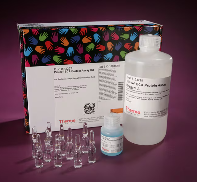

Pierce™ BCA 蛋白检测试剂盒
产品名称: Pierce™ BCA 蛋白检测试剂盒
英文名称: Pierce™ BCA
产品编号: 23227
产品价格: 1919元
产品产地: 进口
品牌商标: null
更新时间: 2025-09-03T13:24:12
使用范围: null
| 规格 | 价格 |
| 500 mL 试剂盒 | 1919.0 |
| 1 Kit (100 mL) | 507.0 |
| 1 L 试剂盒BSA: 2 mg/mL | 3088.0 |
| 1 L 试剂盒BSA: 0.125, 0.25, 0.5, 0.75, 1, 1.5, and 2 mg/mL | 2136.0 |
| 500 mL 试剂盒 BSA: 0.125, 0.25, 0.5, 0.75, 1, 1.5, and 2 mg/mL | 1327.0 |
- 联系人 : 郑立双
- 地址 : 长春市南关区亚泰大街7398号水文化生态园1号楼R307-02
- 邮编 : 130000
- 所在区域 : 吉林省
- 电话 : 186****5058 点击查看
- 传真 : 点击查看
- 邮箱 : tongs_zls@126.com
The Pierce BCA Protein Assay Kit is a high-precision, detergent-compatible protein assay for determination of protein concentration. Pierce BCA reagents provide accurate determination of protein concentration with most sample types encountered in protein research. The Pierce BCA assay can be used to assess yields in whole cell lysates, affinity-column fractions, purified proteins samples, as well as to monitor protein contamination in industrial applications. Compared to most dye-binding methods, the BCA assay is affected much less by protein compositional differences, providing greater concentration accuracy.
Kits are available with or without Pierce Dilution-Free BSA Protein Standards, which are a set of seven pre-diluted BSA standards, packaged in a multichannel pipette-friendly tubestrip. The tubestrip includes a single empty tube that enables users to add their own sample buffer for the purpose of blank subtraction.
The Pierce BCA Protein Assay Kit is a high-precision, detergent-compatible protein assay for determination of protein concentration. Pierce BCA reagents provide accurate determination of protein concentration with most sample types encountered in protein research. The Pierce BCA assay can be used to assess yields in whole cell lysates, affinity-column fractions, purified proteins samples, as well as to monitor protein contamination in industrial applications. Compared to most dye-binding methods, the BCA assay is affected much less by protein compositional differences, providing greater concentration accuracy.
Kit options are available that include Pierce Dilution-Free BSA Protein Standards, which are a set of seven pre-diluted BSA standards, packaged in a multichannel pipette-friendly tubestrip. The tubestrip includes a single empty tube that enables users to add their own sample buffer for the purpose of blank subtraction.
Features of the BCA Protein Assay Kit include:
- Colorimetric—estimate visually or measure with a standard spectrophotometer or plate reader at 562 nm
- Accurate—exhibits half the protein-to-protein variation observed with dye-binding methods (Bradford)
- Compatible—unaffected by typical concentrations of most ionic and non-ionic detergents
- Short assay time—30-min incubation; much easier and four times faster than classical Lowry methods
- Wide assay range—linear working range for BSA of 20 to 2000 μg/mL
- Sensitive—detect down to 5 μg/mL with enhanced protocol
- No more serial dilutions—kit options include Dilution-Free BSA Protein Standards which eliminate the need to perform serial dilutions when generating a standard curve
How the assay works
The BCA Protein Assay combines the well-known reduction of Cu2+ to Cu1+ by protein in an alkaline medium with the highly sensitive and selective colorimetric detection of the cuprous cation (Cu1+) by bicinchoninic acid (BCA). The first step is the chelation of copper with protein in an alkaline environment to form a light blue complex. In this reaction, known as the biuret reaction, peptides containing three or more amino acid residues form a colored chelate complex with cupric ions in an alkaline environment containing sodium potassium tartrate.
In the second step of the color development reaction, BCA reacts with the reduced (cuprous) cation that was formed in step one. The intense purple-colored reaction product results from the chelation of two molecules of BCA with one cuprous ion. The BCA/copper complex is water soluble and exhibits a strong linear absorbance at 562 nm with increasing protein concentrations. The complex is approximately 100 times more sensitive (lower limit of detection) than the pale blue color of the first reaction.
The reaction is strongly influenced by four amino acid residues (cysteine, cystine, tyrosine, and tryptophan) in the amino acid sequence of the protein. However, unlike Coomassie dye-binding methods (i.e., Bradford), the universal peptide backbone also contributes to color formation, helping to minimize variability caused by protein compositional differences.
